
Gout is widely recognized as one of the most agonizing forms of acute inflammatory arthritis known to clinical medicine. Classically presenting as podagra (involvement of the first metatarsophalangeal joint), an acute flare frequently awakens patients from sleep with pain so intense that even the friction of a bedsheet is intolerable.
1. The Superficial Layer: Clinical Neurovascular Aggression
At the gross macroscopic level, the affected joint displays the classical cardinal signs of acute inflammation: extreme calor (heat), rubor (erythema), tumor (edema), and dolor (pain).
- Cutaneous Allodynia & Hyperalgesia: The skin overlying the joint becomes taut, dusky red or violaceous, and exquisitely tender. The inflammatory milieu directly sensitizes cutaneous and articular nociceptors (A-delta and unmyelinated C-fibers), lowering their mechanical threshold so that normally non-noxious stimuli trigger intense nociceptive firing.
- Mechanical Compartment Effect: The first metatarsophalangeal (MTP) joint, midfoot, and ankle are tight, low-capacitance anatomical compartments. As rapid microvascular leakage dumps rich inflammatory exudate and neutrophils into the closed synovial space, intra-articular pressure surges, mechanically stretching the heavily innervated joint capsule and periosteum.
2. The Intermediate Layer: Biophysical & Microenvironmental Drivers
Why does acute gout characteristically attack peripheral acral joints, particularly at night? The answer lies in the biophysics of monosodium urate (MSU) precipitation:
- Temperature-Dependent Solubility: Urate saturation concentration is approximately 6.8 mg/dL at physiological core body temperature (37°C). However, the peripheral temperature of the foot drops to 30–32°C during sleep. This drop sharply shifts the crystallization equilibrium, precipitating sharp, needle-like, negatively birefringent MSU crystals directly into the avascular cartilage and synovium[1].
- Nocturnal Intra-articular Dehydration: During recumbent sleep, water is preferentially reabsorbed from the joint space into the systemic circulation faster than urate can clear, dynamically concentrating synovial fluid and promoting rapid crystallization.
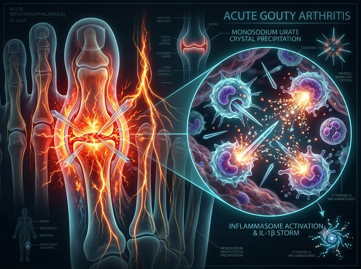
- Physical Morphology of the Crystal: MSU crystals form microscopic, needle-shaped structures with high aspect ratios. Their physical edges pierce and interact with synovial membranes, acting as mechanical irritants and triggering damage-associated danger pathways[2].
3. The Deep Molecular Layer: Phagocytosis and the NLRP3 Inflammasome Storm
The true engine of severe gouty pain occurs at the cellular and immunological interface, mediated by the innate immune system's response to naked uric acid crystals:
A. Opsonization & Phagocytic Rupture
MSU crystals adsorb serum proteins, IgG, and complement components (C3a, C5a) onto their naked surfaces. Resident synovial macrophages and dendritic cells recognize these crystals via Toll-Like Receptors (TLR-2 and TLR-4) and actively engulf them via endocytosis.
Because the rigid crystal surface cannot be digested by lysosomal enzymes, it acts as a molecular needle, destabilizing and lysing the phagolysosome membrane. This rupture releases Cathepsin B into the cytoplasm, signaling cellular stress and massive reactive oxygen species (ROS) formation.
B. Activation of the NLRP3 Inflammasome Complex
Intracellular potassium ($K^+$) efflux coupled with lysosomal disruption activates the NLRP3 inflammasome (NOD-, LRR- and pyrin domain-containing protein 3). Once oligomerized with the adaptor ASC, it recruits and cleaves Pro-Caspase-1 into its catalytically active state, Caspase-1[3].
C. The IL-1β Explosive Cytokine Storm
Caspase-1 rapidly cleaves the inactive cytokine pro-IL-1β into Interleukin-1β (IL-1β), the master molecular mediator of gouty inflammation:
- Chemotactic Influx: IL-1β drives endothelium activation, upregulating selectins, ICAM-1, and chemokines like CXCL8 (IL-8), recruiting hundreds of thousands of polymorphonuclear neutrophils (PMNs) per cubic millimeter of synovial fluid.
- Neutrophil NETosis: Recruited neutrophils engulf crystals, release destructive lysosomal collagenases, protease enzymes, and undergo NETosis (neutrophil extracellular traps), creating a self-amplifying cycle of cellular necrosis.
- Direct Sensitization of Sensory Nerves: IL-1β, along with prostaglandins ($PGE_2$), bradykinin, and substance P, binds directly to receptor terminals on peripheral afferent neurons, inducing robust transcriptional and post-translational hypersensitivity.
Comparative Summary: Why Gout Agony Is Unique
A synthesis of the multi-layered pathological processes driving acute gout flares:
| Layer of Pathology | Primary Trigger | Physiological Consequence | Clinical Manifestation |
|---|---|---|---|
| Superficial | Synovial microvascular leakage into closed joint capsules | High-pressure hydrostatic swelling & nerve compression | Intense allodynia; unable to tolerate even sheets or socks |
| Intermediate | Acral hypothermia (<32°C) & nocturnal fluid shift | Solubility drops below 6.8 mg/dL; sharp needle crystals form | Sudden nocturnal attack onset in the 1st MTP joint (Podagra) |
| Deep (Immunologic) | Macrophage phagolysosome rupture by ingested MSU | NLRP3 assembly & Caspase-1 activation | Rapid systemic pro-inflammatory cytokine surge |
| Molecular Pain | IL-1β release, massive PMN influx & $PGE_2$ cascade | Direct chemical sensitization of peripheral nociceptors | Severe, throbbing, unrelenting acute inflammatory pain |
Understanding this multi-tier inflammatory sequence explains why targeted anti-inflammatory drugs like colchicine (which inhibits microtubule polymerization and neutrophil chemotaxis) and IL-1β antagonists (such as anakinra or canakinumab) provide dramatic symptomatic relief when traditional analgesics fall short[1].